Module 1: Introduction to Microbiology
Introduction
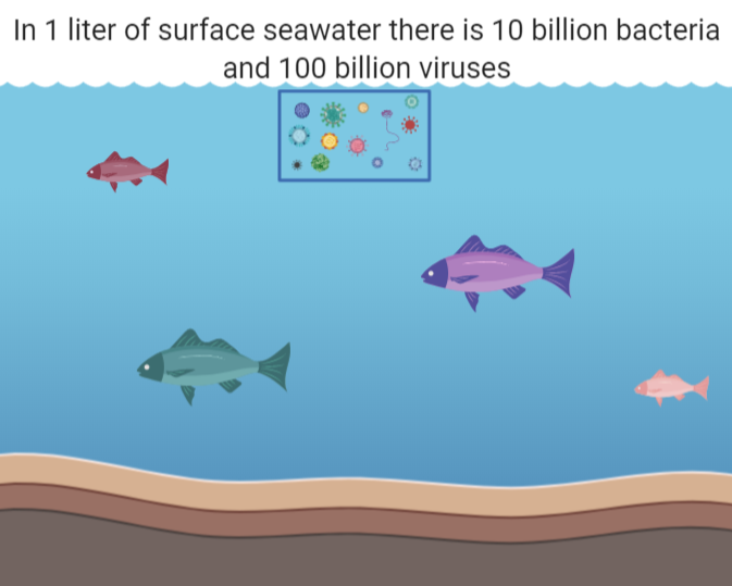
Microorganisms are found almost everywhere on the planet. Due to the incredible amount of microbial diversity, microbes have evolved to live along the deep-sea floor, among soils and roots, and even inside you!
Microbes are essential to life; they serve fundamental roles in our ecosystem and are critically important for nutrient cycling. As primary producers, some microbes generate energy through photosynthesis or chemosynthesis. Nitrogen-fixing microbes remove nitrogen from the atmosphere and transform it into forms useable by other organisms. Some microbes are decomposers; they breakdown organic material and release nutrients into the environment.
Microbes are found across every niche and are essential to life!
Most microbes in the environment are harmless or beneficial, but a small portion of microbes can cause disease. By understanding functions and characteristics of microbes, we may be better able to understand and control the spread of disease.
What are Microbes?
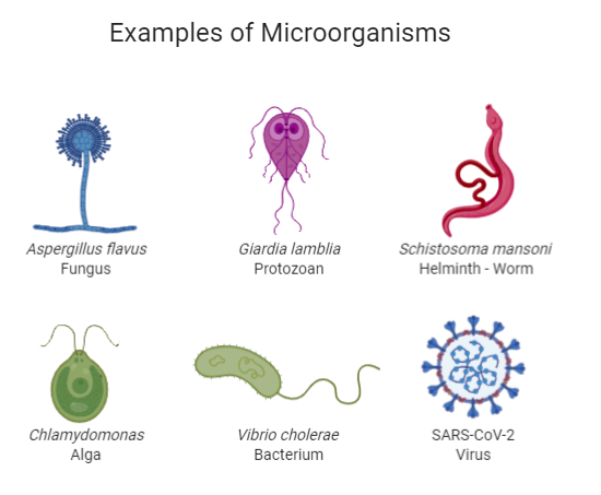
A microorganism, or microbe, is any organism that is too small to be seen by the eyes alone. This includes viruses, bacteria, protozoans, and fungi.
Exercise 1
Exercise
Make a prediction. List the following items from largest to smallest:
Coffee Bean
E. coli Bacteria
Skin Cell
Mitochondria
Viruses
Grain of Salt
Assess your prediction using the Cell Size and Scale Tool.
Solution
A, F, C, D, B, E
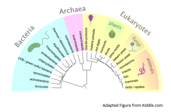
Microorganisms do not belong to only one kingdom; instead, there are a diversity of microorganisms found across each kingdom.
Exercise 2
Exercise
Distinguish between different kinds of microorganisms based on cellular structure. Visit the Cell Anatomy Viewer. Explore the cellular structure of the Animal, Plant, Fungus, and Bacterium. Play the Cell Parts ID Game until you can complete the “Expert Level.” Then, fill in create a chart or diagram to compare the cellular structures, using the terms below.
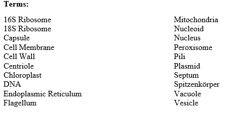
Terms.
Bacteria
Bacteria are single-celled microbes that are found in diverse environments. The more we look, the more places we find them!
Bacteria are categorized by their shape and structure. You have learned that bacterial cells have both a cell membrane and a cell wall. Bacteria are either Gram-positive or Gram-negative based on the structure of their cell wall.

Comparison of Gram-positive bacteria to Gram-negative bacteria.
Question 1
Question
Based on the figure above, what are the key differences between Gram-positive and Gram-negative bacteria?
Solution
Gram-positive bacteria have a thick cell wall that is composed of peptidoglycan. Gram-negative bacteria have a cell wall that is composed of a thin layer of peptidoglycan, surrounded by an outer membrane and lipopolysaccharaides.
There are three basic shapes of bacteria: bacillus (rod-shaped), coccus (spherical), and spirochete (spiral).
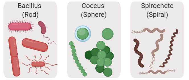
Common shapes of bacteria.
Exercise 3
Exercise
Bacteria are classified as Gram-positive or Gram-negative by the results of the Gram stain method, or Gram’s method. Visit the Gram Stain Virtual Lab. Read the description of the gram stain method and the steps of the method.
The image below is a microscopic image of a culture of Staphylococcus aureus. What is the gram stain of the bacteria? What is the shape of the bacteria?
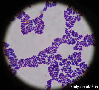
View of Staphylococcus aureus through the microscope, after Gram-stain.
Solution
Staphylococcus aureus is a gram-positive coccus.
Viruses
Viruses are the smallest microbes and require a host cell to survive. In order to replicate, a virus must infect a host cell. These host cells may be cells from humans, plants, animals, or other microorganisms.
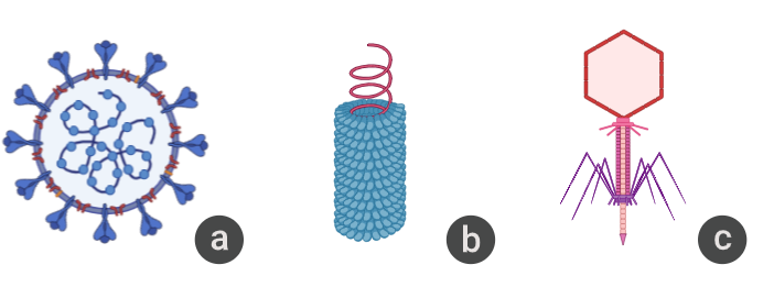
Examples of viruses. (a) The SARS-CoV02 virus infects and replicates inside human and animal cells, (b) The Tobacco Mosaic Virus infects and replicates inside a wide range of plants, especially tobacco, (c) The bacteriophage virus infects and replicates inside bacterial cell hosts.
Examples of viruses. (a) The SARS-CoV02 virus infects and replicates inside human and animal cells, (b) The Tobacco Mosaic Virus infects and replicates inside a wide range of plants, especially tobacco, (c) The bacteriophage virus infects and replicates inside bacterial cell hosts.
Watch a virus in action, here! In this animation, a bacteriophage virus infects an E. coli host cell. The step-by-step infection is explained here.
Are viruses alive?
This is a common debate, even among scientists!
Viruses are acellular, meaning that they do not have a cellular structure. Viruses do have genetic material that is protected by a protein coat.
In order to reproduce, viruses must infect and hijack their hosts’ cell or cells in order to replicate their own genetic material. Viruses use the hosts’ cellular machinery, metabolism, and nutrition in order to replicate.
Some viruses may respond to conditions in the host’s cell. For example, if a bacteriophage virus enters a host cell, the virus may enter a stage of virulent infection, known as the lytic cycle. During the lytic cycle, the virus will hijack the hosts’ machinery immediately. The virus will replicate and then will lyse, or burst, the host’s cell.
In contrast, a bacteriophage virus may ‘decide’ to remain in a dormant inside the host’s cell. In this case, the virus may enter the lysogenic cycle, where the viral DNA may replicate but will not burst the host’s cell. If the host cell encounters an environmental stressor, the virus may then transition into the lytic cycle, replicating and lysing the host cell.

Lytic Vs. Lysogenic Cycles of Bacteriophage
Virus Structure
All viruses are composed of genetic material and a protein coat, but viruses may take many different forms and structures. Viruses are classified according to their composition and structure, including their genetic material, their physical shape, and their outer coating.
Genetic Material. Viruses may use DNA or RNA for their genetic material. This material may be double-stranded or single-stranded.
Physical Shape. Viruses are classified according to their geometric shape as either helical, polyhedral, spherical, or complex.
Outer Coating. All viruses have a protein coat. Enveloped viruses also have a lipid bilayer membrane and glycoproteins. Non-enveloped viruses have only a protein coat, but, non-enveloped viruses are more persistent, or hardy, in the environment.
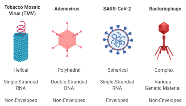
Comparison of four different viruses, based on shape, genetic material, and outer coating.
Wrap-Up Exercise
Exercise 4
Exercise
How do viruses compare to other microorganisms? Generate a table to compare viruses to other microorganisms based on size, genetic material, structure, and reproduction.
Solution
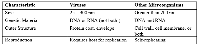
Comparison between viruses and other microorganisms.